-
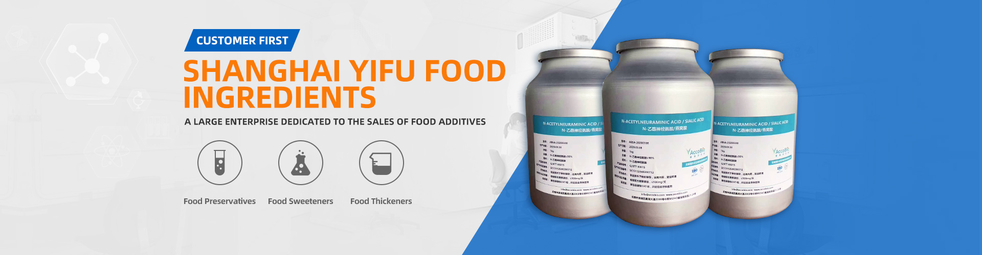
Tel:15618093168
|
Email:2307176274@qq.com
Wound healing is a complex physiological process involving inflammatory responses, cell proliferation, and tissue remodeling, influenced by multiple factors such as nutritional status, cytokine regulation, and oxidative stress. L-arginine, a semi-essential amino acid in humans, not only serves as a key building block for protein synthesis but also generates bioactive molecules (e.g., nitric oxide [NO], polyamines) through metabolic pathways, exerting regulatory effects across multiple stages of wound healing. Recent studies have shown that exogenous supplementation of L-arginine significantly accelerates the healing of chronic non-healing wounds (e.g., diabetic foot ulcers, pressure sores) and acute trauma. Its mechanisms of action are closely associated with improving the local microenvironment, regulating cell function, and inhibiting inflammatory imbalance. Starting from the physiological stages of wound healing, this article systematically analyzes the promotional effects and molecular mechanisms of L-arginine, providing a theoretical reference for clinical wound management.
I. Adaptation to Physiological Stages of Wound Healing: Whole-Process Regulation from Inflammation to Remodeling
Wound healing typically proceeds through three stages: the inflammatory phase (0–3 days), proliferative phase (3–14 days), and remodeling phase (2 weeks to several months). Each stage has distinct cellular activities and microenvironmental requirements. L-arginine exerts targeted effects at each stage through its metabolic properties, forming a "whole-process" regulatory pattern:
(I) Inflammatory Phase: Regulating Inflammatory Responses and Alleviating Oxidative Damage
The inflammatory phase initiates wound healing, primarily relying on neutrophils and macrophages to clear necrotic tissue and pathogens. However, excessive inflammation or oxidative stress delays healing. The core role of L-arginine in this phase is to "balance inflammation and antioxidant defense":
On one hand, L-arginine is catalyzed by nitric oxide synthase (NOS) to produce NO. NO dilates local blood vessels in the wound, increasing blood flow, oxygen, and nutrient supply. It also inhibits excessive platelet aggregation to prevent thrombus formation (thrombi block local blood supply, leading to tissue hypoxia). Additionally, as a signaling molecule, NO regulates macrophage activation—promoting the differentiation of anti-inflammatory M2-type macrophages and inhibiting the overproliferation of pro-inflammatory M1-type macrophages. This reduces the release of pro-inflammatory factors such as tumor necrosis factor-α (TNF-α) and interleukin-6 (IL-6), preventing prolonged inflammation (e.g., an overabundance of M1-type macrophages in chronic wounds continuously releases inflammatory factors, hindering the initiation of healing).
Furthermore, L-arginine enhances the synthesis of glutathione (GSH), boosting the local antioxidant capacity of the wound. GSH is a critical antioxidant that scavenges reactive oxygen species (ROS) generated during the inflammatory phase, reducing ROS-induced damage to vascular endothelial cells and fibroblasts (excessive ROS causes cell apoptosis and impairs wound tissue integrity). Experiments show that in mice supplemented with L-arginine, wound ROS levels decrease by 40% and vascular endothelial cell apoptosis rate drops by 35% during the inflammatory phase, laying a foundation for the subsequent proliferative phase.
(II) Proliferative Phase: Promoting Cell Proliferation, Matrix Synthesis, and Accelerating Tissue Repair
The proliferative phase is critical for "filling wound defects," relying on fibroblast proliferation, angiogenesis, epithelial cell migration, and collagen synthesis. L-arginine drives this phase through "metabolic substrates" and "signal regulation":
Fibroblast proliferation and collagen synthesis: Fibroblasts are the primary cells responsible for collagen synthesis, and L-arginine is an essential precursor for collagen (approximately 30% of amino acids in collagen molecules are proline and glycine, and L-arginine can be metabolically converted into proline precursors). Meanwhile, NO generated from L-arginine activates the cyclic guanosine monophosphate (cGMP) signaling pathway in fibroblasts, promoting the cell cycle transition from the G1 phase to the S phase and accelerating fibroblast proliferation (in vitro experiments show that the proliferation rate of fibroblasts in the L-arginine treatment group is 60% higher than that in the control group). Additionally, NO regulates collagen cross-linking, enhancing the strength of collagen fibers (uncross-linked collagen is easily degraded, leading to poor wound healing).
Angiogenesis: Angiogenesis provides oxygen and nutrients to cells during the proliferative phase. L-arginine promotes angiogenesis through two pathways: 1) NO activates the expression of vascular endothelial growth factor (VEGF), a specific growth factor for vascular endothelial cells that induces endothelial cell migration and tubular structure formation; 2) L-arginine is metabolized by arginase to produce ornithine, which is further converted into polyamines (putrescine, spermidine). Polyamines stabilize the DNA of vascular endothelial cells, promote cell division, and accelerate capillary network construction (polyamine deficiency causes stagnation of vascular endothelial cell proliferation and impairs angiogenesis).
Epithelial cell migration: Epithelial cell migration to cover the wound surface marks "wound closure." NO generated from L-arginine reduces the adhesion between epithelial cells, promoting their migration toward the wound center while inhibiting epithelial cell apoptosis (apoptosis disrupts epithelial layer repair). Clinical studies show that in burn patients treated with topical L-arginine gel, the migration rate of epithelial cells increases by 50%, and wound closure time is shortened by 3 days compared to the control group.
(III) Remodeling Phase: Optimizing Collagen Remodeling and Enhancing Wound Strength
The core of the remodeling phase is the "ordered remodeling" of collagen—converting disordered collagen fibers synthesized during the proliferative phase into mature, orderly arranged collagen bundles, while clearing excess cells and matrix to enhance wound tensile strength. L-arginine plays a role in "regulating remodeling balance" during this phase:
NO generated from L-arginine activates matrix metalloproteinases (MMPs), key enzymes that degrade excess collagen. MMPs clear uncross-linked collagen residues and damaged matrix from the proliferative phase, preventing excessive collagen accumulation and scar hyperplasia (collagen in scar tissue is disorderly arranged, with tensile strength only 70% of normal skin). Meanwhile, NO inhibits excessive MMP activation (excessive MMPs degrade normal collagen, leading to fragile wounds), maintaining a "synthesis-degradation" balance.
Additionally, L-arginine promotes the differentiation of fibroblasts into myofibroblasts. Myofibroblasts reduce wound area through contraction and secrete mature type I collagen (the main type of skin collagen, accounting for approximately 80% of total collagen with high tensile strength), improving wound repair quality. Experiments show that in rats supplemented with L-arginine, the proportion of type I collagen in wounds increases from 65% to 85% during the remodeling phase, and wound tensile strength improves by 40%, approaching the level of normal skin.
II. Core Molecular Mechanisms for Promoting Wound Healing: Multi-Pathway Synergistic Regulation
The promotional effect of L-arginine on wound healing essentially involves bioactive molecules generated through its metabolic pathways (NOS pathway, arginase pathway) regulating intracellular signaling pathways and gene expression. These mechanisms can be summarized into three core pathways:
(I) NO-cGMP Signaling Pathway: The Core Pathway Regulating Vascular and Cellular Function
NO, produced by L-arginine under NOS catalysis, is a key signaling molecule for wound healing, primarily exerting its effects through the NO-cGMP signaling pathway:
After entering cells, NO activates soluble guanylate cyclase (sGC), which catalyzes the conversion of guanosine triphosphate (GTP) to cGMP. As a second messenger, cGMP activates protein kinase G (PKG), which phosphorylates downstream target proteins to exert multiple effects:
Phosphorylates the myosin light chain of vascular smooth muscle cells, causing smooth muscle relaxation, vasodilation, and increased local blood supply to the wound.
Phosphorylates cyclin D1 in fibroblasts, promoting cell cycle progression and accelerating proliferation.
Phosphorylates VEGF receptors on vascular endothelial cells, enhancing VEGF signal transduction and promoting angiogenesis.
Additionally, cGMP inhibits the NF-κB signaling pathway in inflammatory cells (NF-κB is a core transcription factor for pro-inflammatory factor expression), reducing the release of TNF-α and IL-6 and alleviating inflammatory damage. When NOS activity is inhibited (e.g., L-arginine deficiency), NO production decreases, cGMP levels drop, and activation of the above pathways is blocked, significantly delaying wound healing.
(II) Arginase-Polyamine Pathway: Metabolic Support for Cell Proliferation and Matrix Synthesis
L-arginine is catalyzed by arginase to produce ornithine, which is converted into polyamines (putrescine, spermidine) under the action of ornithine decarboxylase (ODC). Polyamines are essential for cell proliferation and matrix synthesis, with mechanisms including:
Stabilizing DNA and RNA structures: Polyamines bind to nucleic acid molecules, maintaining the stability of the DNA double helix structure, preventing DNA damage during cell division, and promoting RNA transcription and protein synthesis (e.g., collagen, VEGF).
Regulating cytoskeleton reorganization: Polyamines bind to tubulin, promoting microtubule assembly—microtubules are critical structures for cell migration and division (fibroblast migration depends on microtubule-mediated cytoskeleton reorganization).
Inhibiting cell apoptosis: Polyamines activate the expression of the anti-apoptotic protein Bcl-2 and inhibit the activity of the pro-apoptotic protein Bax, reducing apoptosis of fibroblasts and vascular endothelial cells (apoptosis leads to insufficient cell numbers during the proliferative phase, delaying wound repair). Studies indicate that in mice with inhibited arginase activity (blocking polyamine production), wound fibroblast proliferation rate decreases by 50%, collagen synthesis reduces by 45%, and healing time doubles.
(III) Oxidative Stress Regulation: A Protective Mechanism for Improving the Wound Microenvironment
Excessive oxidative stress (excess ROS) in the wound is a major cause of delayed healing. L-arginine improves the microenvironment through a dual mechanism of "scavenging ROS + enhancing antioxidant enzyme activity":
L-arginine serves as a precursor for glutathione (GSH) synthesis (GSH synthesis requires cysteine, and L-arginine promotes cysteine production through metabolism). GSH directly scavenges ROS (e.g., superoxide anions, hydrogen peroxide), reducing ROS-induced oxidative damage to cells.
Additionally, NO generated from L-arginine activates the activity of antioxidant enzymes such as superoxide dismutase (SOD) and catalase (CAT). SOD converts superoxide anions into hydrogen peroxide, and CAT further decomposes hydrogen peroxide into water, forming a "ROS scavenging chain."
For chronic non-healing wounds (e.g., diabetic foot ulcers), patients often experience excessive oxidative stress (hyperglycemia induces massive ROS production). Supplementation with L-arginine significantly reduces wound ROS levels and enhances antioxidant enzyme activity—clinical studies show that after 8 weeks of oral L-arginine supplementation (3g daily) in patients with diabetic foot ulcers, local wound SOD activity increases by 60%, ROS levels decrease by 55%, and ulcer healing rate rises from 30% to 65%.
III. Application Forms and Clinical Adaptation: Strategies for Different Wound Types
Based on the above mechanisms, the application of L-arginine should align with wound types (acute/chronic, superficial/deep) to select appropriate supplementation forms and maximize promotional effects:
Acute trauma (e.g., surgical incisions, superficial burns): Oral supplementation (2–3g L-arginine daily) combined with topical L-arginine gel (5%–10% concentration) is recommended. Oral supplementation increases systemic amino acid levels to provide substrates for wound healing, while topical application acts directly on wound tissue, rapidly increasing NO concentration and accelerating inflammation resolution and epithelial repair.
Chronic non-healing wounds (e.g., diabetic foot ulcers, pressure sores): Patients often have insufficient local blood supply and prolonged inflammation, so a combined "oral + local injection" regimen is suggested—3–4g oral daily, combined with twice-weekly local injections of L-arginine solution (10% concentration). Injections directly improve the local microenvironment and promote angiogenesis.
Malnutrition-related wounds (e.g., wounds in elderly patients, post-tumor surgery): Patients have insufficient endogenous amino acid reserves, so oral dosage should be increased (4–5g daily) and combined with protein supplementation (e.g., whey protein). This prevents L-arginine from being prioritized for basic protein synthesis, ensuring its allocation to wound metabolic pathways (NOS, arginase pathways).
L-arginine exerts multiple effects—"balancing inflammation, promoting proliferation, and optimizing remodeling"—across the inflammatory, proliferative, and remodeling phases of wound healing through the NO-cGMP signaling pathway, arginase-polyamine pathway, and oxidative stress regulation. It is particularly effective in promoting the healing of chronic non-healing wounds and acute trauma. The core of its molecular mechanism lies in bioactive molecules (NO, polyamines) generated through metabolism, which precisely regulate cell function and the microenvironment, compensating for "substrate deficiency" and "signal imbalance" during wound healing.
Future research can focus on three areas: 1) developing targeted delivery systems for L-arginine (e.g., nanocarriers) to achieve precise local release in wounds and reduce systemic side effects; 2) exploring its synergistic effects with other pro-healing factors (e.g., VEGF, growth hormone) to further improve healing efficiency; 3) optimizing dosage regimens for special populations (e.g., patients with severe diabetes, immunodeficiency) to provide more precise theoretical support for personalized clinical wound management.